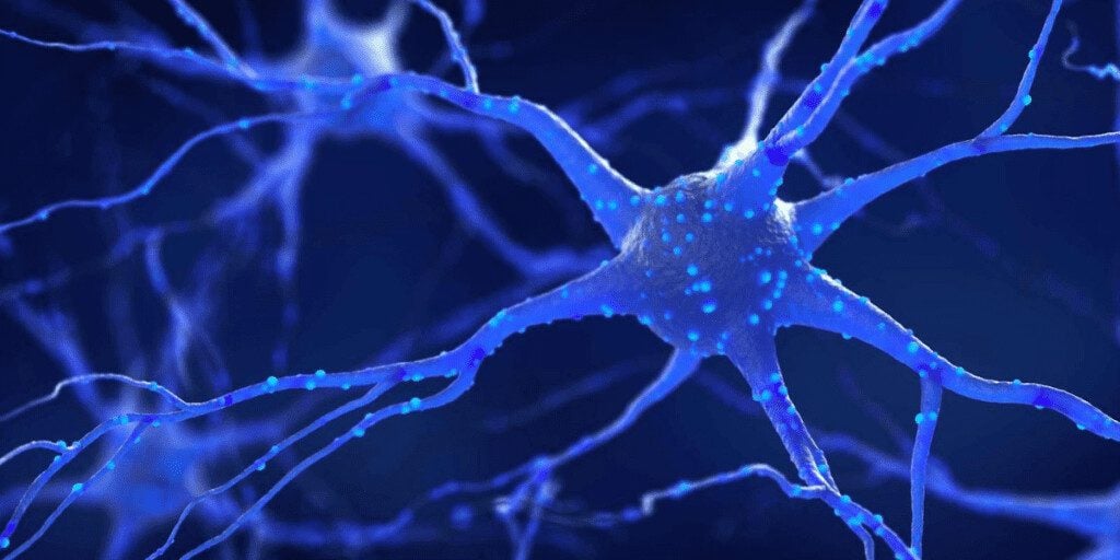
When it comes to the progression of ALS, the causes of cell damage are unclear. New research suggests that the gene damage responsible for neurological breakdown may also trigger an overabundance of stress granules and toxins in patients with ALS. 1
The role of physical or environmental stress in the progress of ALS has long been recognized, but researchers are beginning to understand how stress affects the progression on a cellular and subcellular level. The impact of stress granules and the build-up of protein within the cells is a critical factor in ALS.
What are Stress Granules?
In response to stress, such as heat, cold, or excessive glucose, the body’s cells produce biochemical proteins called “stress granules.” These ribonucleoprotein complexes are formed when the cell slows down processes after a stress incident and bundles the waste products for disposal. This process, called “ubiquitination” is essential for cell health. “Ubiquitin” is the protein added to the cell to mark waste products for removal.
Until recently, the exact mechanism by which stress granules were reformed after stress, and what their removal or lack of removal did to the cell, was unclear. A detailed study of the mechanisms of cell biology, though, has now examined the impact of the formation and removal of the granules. 2
What Do Stress Granules Do?
Stress granules form in all bodies when the body is faced with cellular stress and the need to move protein rapidly from one part of the body to another. Stress granules collect RNA and mRNA and prevent the breakdown of protein, ensuring that it is available for transport through the cells.
What Do Stress Granules Contain?
Stress granules are protein complexes that contain RNA-binding proteins, mRNA, and translation factors. By binding these in a cytoplasmic bundle, the stress granules prevent RNA/mRNA transcription until the stressor has passed. Once the stress is over, the stress granules are supposed to disassemble, and mRNA transcription can resume.
What Role Does a Stress Granule’s Central Protein Play?
There are a number of proteins in stress granules, but the central protein G3BP1 is essential for the disassembly process.3 This protein has been shown to be important when cells are subjected to heat shock, but not other forms of cellular shock.
When stress granules break down, the RNA/mRNA functions return to normal, and cell growth resumes. When stress granules do not break down, on the other hand, protein continues to build up in the cells, and normal RNA transcription does not occur. This may be one cause of the neurological degeneration and downstream cell loss associated with ALS.

Stress Granules and TDP-43 Regulation
TDP-43 is a DNA-binding protein similar to the RNA-binding proteins mentioned. TDP-43 has been implicated in causing protein formations in nerve bundles in ALS patients.4 This protein may also be involved in the breakdown of stress granules or the failure to break them down.
TDP-43 is utilized during cellular stress to assist in the sequestration and transport of mRNA within the cell.5 One study suggested that TDP-43 had a greater impact on the breakdown of stress granules in oxidative stress compared to the effect of G3BP1, which was critical for heat stress. It may be that multiple types of protein are needed for complete stress granule breakdown.
Stress Granules and Neurodegeneration
Incomplete removal of protein—sometimes called “misfolding” or “aggregation”—has been long recognized as a cause of neurodegenerative disorders.6 Researchers are now learning how stress causes proteins to accumulate in the cells and why that is so damaging to the body.
One theory has been that these misfolded proteins become “prions” (proteinaceous infectious particles). Stress granules strongly resemble prion-like agents like those implicated in so-called “mad-cow disease” and Creutzfeldt-Jakob Disease, at least in terms of their effects. However, no transmissibility has been shown with stress granules or any other misfolded proteins.
The transportation of proteins and the mechanics of folding and disassembling them for removal are the functions of the valosin-containing protein (VCP) gene. This VCP gene is responsible for creating an enzyme that handles fusing and breaking down cells and cell membranes. Not surprisingly, variations in the VCP gene have also been implicated in ALS and similar disorders.7
Environmental Stressors and ALS
It is important to note that the type of stress under discussion here is not emotional or psychological stress. Mental stress plays a role in the health and well-being of an ALS patient but may have little impact on their physical and cellular health. The stressors which affect the formation of stress granules and may inhibit their breakdown are environmental.8
Environmental factors that increase the risks for cellular stress:
- Smoking. Physical damage to the lungs and blood vessels may increase damage on a cellular level and increase the potential for harmful genetic mutation.
- Military service. This service may be associated with a higher risk of exposure to environmental toxins.
- Lead exposure or pesticide exposure. The potential impact of these substances on cellular reproduction are well known.
- Electromagnetic radiation has not been well-studied in relation to ALS but cannot be ruled out. Other types of radiation need to be examined for possible cell damage.
- Male sex. It is not yet clear if there is a genetic component to ALS, but it is known that males are more susceptible than females.
These types of environmental stressors place a strain on the cells themselves and initiate the cellular mechanisms that create stress granules. Hot, cold, and toxic environments are the types of stress most commonly encountered by ALS patients. It may be that emotional stress increases the damage caused by environmental stress, but this needs to be studied more closely.

The Impact of Stress Granules Research
Research into stress granules, their formation, and the role they play in the progression of the disease continues. The impact of cellular stress and the role of genetic damage continues to be felt by patients and their families every day. Researchers and the foundations that support them, though, also continue to work every day on ways to identify the disease and develop ways to combat the disorder and prevent it from progressing.
Frequently Asked Questions:
What is the difference between P bodies and stress granules?
The key difference between P bodies and stress granules is that the former assembles around key enzymes of cytoplasmic RNA degradation, while the latter comes together around the components of the translation machinery.
What proteins are in stress granules?
There is a combination of proteins in stress granules, including mRNA and RNA-binding proteins, plus 40S ribosomal subunits.
How do stress granules disassemble?
Through a process involving RNA leaving that causes structural instability and a breakdown into smaller core structures.
SOURCES
1. Maxwell, B. A., Gwon, Y., Mishra, A., Peng, J., Nakamura, H., Zhang, K., Kim, H. J., & Taylor, J. P. (2021). Ubiquitination is essential for recovery of cellular activities after heat shock. Science, 372(6549). https://doi.org/10.1126/science.abc3593
2. Li, Y. R. (2013, April 29). Stress granules as crucibles of ALS pathogenesis | Journal of Cell Biology | Rockefeller University Press. Journal of Cell Biology. Retrieved July 6, 2022, from https://rupress.org/jcb/article/201/3/361/37270/Stress-granules-as-crucibles-of-ALS
3. Gwon, Y., Maxwell, B. A., Kolaitis, R. M., Zhang, P., Kim, H. J., & Taylor, J. P. (2021). Ubiquitination of G3BP1 mediates stress granule disassembly in a context-specific manner. Science, 372(6549). https://doi.org/10.1126/science.abf6548
4. Kurashige, T., MD PhD. (2022, May 23). TDP-43 Accumulation Within Intramuscular Nerve Bundles of Patients With Amyotrophic Lateral Sclerosis. Amyotrophic Lateral Sclerosis | JAMA Neurology | JAMA Network. Retrieved July 6, 2022, from https://jamanetwork.com/journals/jamaneurology/fullarticle/2792745
5. Khalfallah, Y. (2018, May 15). TDP-43 regulation of stress granule dynamics in neurodegenerative disease-relevant cell types. Nature. Retrieved July 6, 2022, from https://www.nature.com/articles/s41598-018-25767-0?error=cookies_not_supported&code=2eea638c-ffe6-4fe9-a031-2583653f8f5d
6. Soto, C., PhD. (2008, February 1). Protein Misfolding and Neurodegeneration. Neurology | JAMA Neurology | JAMA Network. Retrieved July 6, 2022, from https://jamanetwork.com/journals/jamaneurology/fullarticle/795151
7. VCP gene: MedlinePlus Genetics. (n.d.). Medline. Retrieved July 6, 2022, from https://medlineplus.gov/genetics/gene/vcp/#resources
8. Oskarsson, B., Horton, D. K., & Mitsumoto, H. (2015). Potential Environmental Factors in Amyotrophic Lateral Sclerosis. Neurologic Clinics, 33(4), 877–888. https://doi.org/10.1016/j.ncl.2015.07.009


